


Cathodoluminescence Imaging and Spectral Detection System——Rainbow

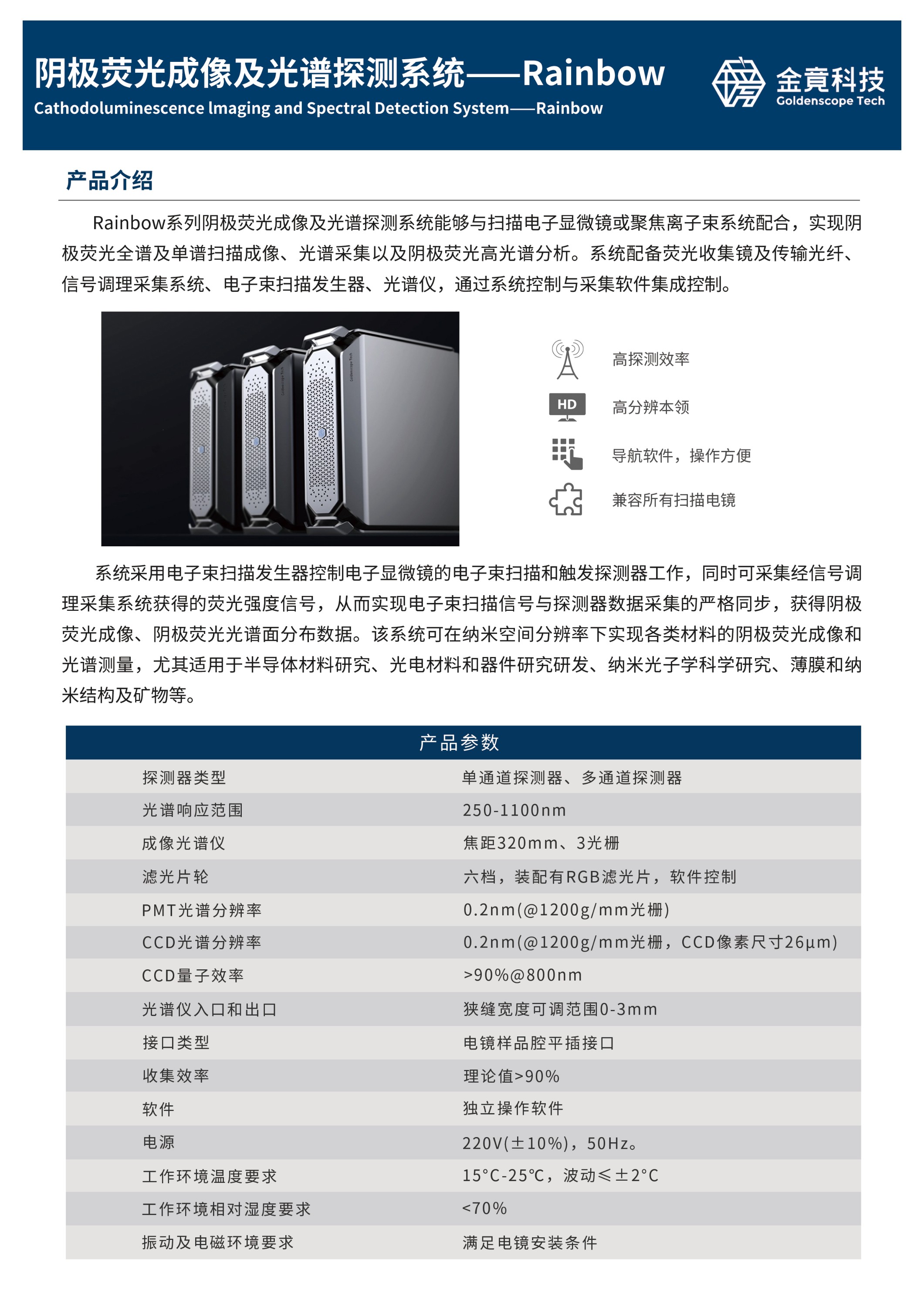
The Rainbow Series Cathodoluminescence Imaging and Spectral Detection System can be integrated with scanning electron microscopes or focused ion beam systems to achieve full-spectrum and single-spectrum scanning imaging, spectral acquisition, and hyperspectral analysis of cathodoluminescence. The system is equipped with a fluorescence collection mirror, transmission optical fibers, a signal conditioning and acquisition system, an electron beam scanning generator, and a spectrometer, all controlled through integrated system control and acquisition software.
The system utilizes the electron beam scanning generator to control the electron beam scanning of the electron microscope and to trigger the detectors. Simultaneously, it can collect fluorescence intensity signals obtained through the signal conditioning and acquisition system, ensuring strict synchronization between the electron beam scanning signal and the detector data acquisition. This allows for the acquisition of cathodoluminescence imaging and cathodoluminescence spectral surface distribution data. The system enables cathodoluminescence imaging and spectral measurements of various materials at nanoscale spatial resolution. It is particularly well-suited for research in semiconductor materials, optoelectronic materials and devices, nanophotonics, thin films, nanostructures, and minerals.


(1)CL Functionality: Monochromatic, full-color, and RGB CL imaging. Capable of acquiring point, line, and surface CL spectra. Features a wavelength monochromator imaging function and uses a PMT detector for rapid imaging at user-selected wavelengths.
(2)Spectrometer Design: C-T (Czerny-Turner) design with super toroidal imaging calibration, providing achromatic full-spectrum and broad spectral coverage.
(3)High-Performance Ellipsoidal Mirror Collection: High CL collection efficiency with a total efficiency of up to 90% (Lambertian source). Detector types include single-channel (PMT) and multi-channel (CCD) detectors.
(4)Spectral Response Range: 200-1100 nm.
(5)Enhanced Spectrometer: Features a 320 mm focal length and three gratings, utilizing a 68×68 large-area grating to improve light flux detection capability. The spectrometer coupling module precisely adapts and matches the CL collimated beam with the spectrometer's F-number (aperture), F=4.2.
(6)CL Intensity Imaging: Uses a high-speed PMT detector with a wavelength response range of 160-900 nm.
(7)CCD Spectral Resolution: 0.2 nm (with a 1200 g/mm grating, CCD pixel size of 15 μm); PMT resolution: 0.2 nm (with a 1200 g/mm grating).
(8)Professional CL Spectral Acquisition: Operates with standalone software for easy data acquisition and analysis. The open-source software simplifies the entire CL imaging process. The system supports point scanning, line scanning, and surface scanning with full-spectrum, single-spectrum, and hyperspectral imaging capabilities, all with a fully Chinese-language user interface.


Professional Systematic Training


Rich Application Experience


Timely And Efficient Response


Excellent Service Experience


GOLDENSCOPE TECH GOLDENSCOPE TECH GOLDENSCOPE TECH GOLDENSCOPE TECH GOLDENSCOPE TECH GOLDENSCOPE TECH GOLDENSCOPE TECH GOLDENSCOPE TECH GOLDENSCOPE TECH GOLDENSCOPE TECH


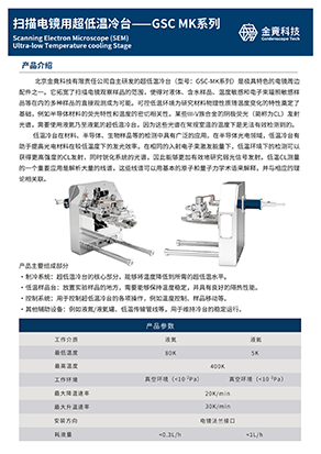
 Product Introduction
 Technical Specifications
 Download Center
 Service Support